

|
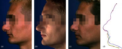
Patient Case no. 1 The first patient suffers from a so-called short face syndrome which is a retrodisplacement of the maxilla and mandible (upper and lower jaw) in combination with a deep bite because of a predominantly horizontal growth pattern of the bases of the jaws in relation to the skull base (see figure a). |
|
The correspondence of simulation (figure b) and real surgery (figure c) is exceptional. The profile lines are given in figure (d), where blue, green and red represent pre-surgical, simulated and post-surgical situation respectively. Minor deviations can only be observed in the region of the mouth. |

|
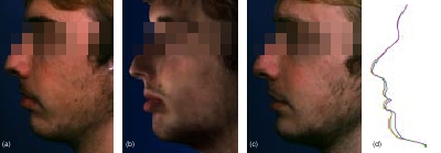
Patient Case no. 2 The second case shows a complex facial asymmetry due to a chronic disease of the facial bones in the left midface (fibrous dysplasia, figure a). Overgrowth of the left half of the lower jaw, the left cheekbone and the bones surrounding the eye sockets (periorbital bones) led to a severe transversal deviation of the occlusal plane. |
|
The simulation of a case of such complexity is demanding due to the correction of asymmetries as well as because of the rotations and the genioplasty introduced during surgery. Although, as can be seen in the profile lines, the correspondence at the chin and lips is satisfactory. |

|
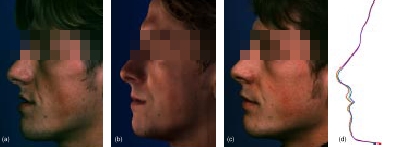
Patient Case no. 3 The fourth case shows a patient suffering from excessive vertical growth of the retrodisplaced maxilla. In this case, only the molar teeth are in contact. The incisor teeth are too far behind and therefore cannot touch their antagonists in the mandible. In addition, the lower jaw is overprojected to a certain extent (figure a). |
|
The surgical procedure only introduced a very small displacement field. Although the resulting contour lines are acceptable the accurate simulation of such small bone movements lies beyond the resolution bounds of the system. |

|
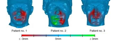
Error Visualization In addition to the profile views shown in the figures above the figure to the left depicts the results of a quantitative error analysis in terms of the distance between predicted and post-surgical surface. The colors are computed from the radial distance between the two surfaces projected onto a local normal. |
|
Positive values indicate the predicted surface lying in front of the post-surgical whereas red colors illustrate the contrary. Errors at the side of the nose common to all cases must be attributed to the radial measuring set-up which tends to overemphasize errors at places where the surface happens to be of nearly the same orientation as the radial direction of measurement. Further, errors at the cheekbones are due to swelling. The case of the second patient shows that asymmetric corrections are very demanding and therefore cannot be predicted completely accurately. |